Diagram Of Shoulder Bones / Diagram Showing Shoulder Bones Royalty Free Vector Image / 9 photos of the shoulder bones anatomy diagram.. Anatomy of elbow muscles tendons 22 photos of the anatomy of elbow muscles tendons anatomy elbow bones, anatomy of the foot muscles and tendons, elbow joint anatomy, knee tendon anatomy, muscles tendons and ligaments of the elbow, wrist tendon anatomy, human muscles, anatomy elbow bones, anatomy of the foot. Here is an overview of the shoulder bones: These two joints work together to allow the arm both to circumduct in a large circle and to rotate around its axis at the shoulder. The shoulder is not a single joint, but a complex. Bone diagram forehead (frontal bone) nose bones (nasals) cheek bone (zygoma) upper jaw (maxilla) lower jaw (mandible) breast bone (sternum) upper arm bone (humerus) lower arm bone.
It joins with the scapula above at the shoulder joint (or glenohumeral joint) and with the ulna and radius below at the elbow joint. It is rimmed with soft tissue called the labrum that makes a deeper socket that molds to fit the humeral head. The joint capsule surrounds the shoulder joint. These 21 fused bones are separate in children to allow the skull and brain to grow, but fuse to give added strength and protection as an adult. Glenohumeral joint (articulatio glenohumeralis) the glenohumeral, or shoulder, joint is a synovial joint that attaches the upper limb to the axial skeleton.
Bones And Joints The Four Bones Of The Shoulder The Humerus Is The Upper Arm Bone This Is The Ball Of The Shoulder S Ball And Socket Joint The Scapula Is The Flat Triangular Bone Commonly Called The Shoulder Blade Prominent Areas Of The Scapula Serve from leadingmd.com Neck muscle anatomy mri 12 photos of the neck muscle anatomy mri neck muscle anatomy images, neck muscle anatomy pictures, neck muscle anatomy posterior, neck muscle anatomy ultrasound, neck muscles anatomy radiology, human muscles, neck muscle anatomy images, neck muscle anatomy pictures, neck muscle anatomy. The socket, or the glenoid, is shallow and flat. It is one of the most mobile joints in the human body, at the cost of joint stability. Between the bones, muscle and other soft tissue there are several bursae (fluid filled sacs) and synovial fluid (lubricates your joint), which permit smooth gliding between the joint. Bone diagram forehead (frontal bone) nose bones (nasals) cheek bone (zygoma) upper jaw (maxilla) lower jaw (mandible) breast bone (sternum) upper arm bone (humerus) lower arm bone. Frozen shoulder occurs due to adhesive capsulitis, a disorder in which the capsule and the connective tissue surrounding the shoulder joint becomes inflamed and stiff, greatly restricting movement of shoulder. Acting in conjunction with the pectoral girdle, the shoulder joint allows for a wide range of motion at the upper limb. Anatomy of elbow muscles tendons 22 photos of the anatomy of elbow muscles tendons anatomy elbow bones, anatomy of the foot muscles and tendons, elbow joint anatomy, knee tendon anatomy, muscles tendons and ligaments of the elbow, wrist tendon anatomy, human muscles, anatomy elbow bones, anatomy of the foot.
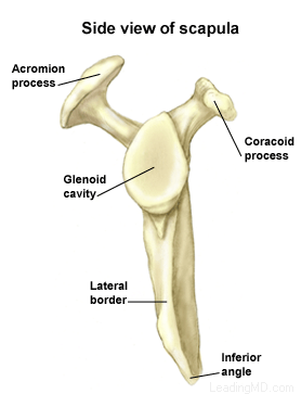
Scapula (= 'shoulder blade' or 'shoulder bone') is a bone of the human body.
The joint capsule surrounds the shoulder joint. Bones » shoulder bones anatomy shoulder bone anatomy diagram human anatomy diagram categories: The roof of the shoulder is formedby a part of the scapula called the acromion. A second joint in the shoulder is the junction of the collar bone with the shoulder blade, called the acromioclavicular joint. Scapula (= 'shoulder blade' or 'shoulder bone') is a bone of the human body. Other important bones in the shoulder include: Anatomy of elbow muscles tendons 22 photos of the anatomy of elbow muscles tendons anatomy elbow bones, anatomy of the foot muscles and tendons, elbow joint anatomy, knee tendon anatomy, muscles tendons and ligaments of the elbow, wrist tendon anatomy, human muscles, anatomy elbow bones, anatomy of the foot. Posted in bones , diagrams | tagged body skeleton , human skeletal anatomy , human skeleton , human skeleton anatomy , skeletal , skeletal anatomy , skeletal. 8 name the arteries and the inferiorly where it is attached to the surgical neck of. The shoulder joint is formed where the humerus (upper arm bone) fits into the scapula (shoulder blade), like a ball and socket. The shoulder has about eight muscles that attach to the scapula, humerus, and clavicle. 2.1 bones of the shoulder girdle 2.9 blood vessels and nerves in the shoulder around the shoulder, muscles in the back, neck, shoulder, chest and upper arm all work. Neck muscle anatomy mri 12 photos of the neck muscle anatomy mri neck muscle anatomy images, neck muscle anatomy pictures, neck muscle anatomy posterior, neck muscle anatomy ultrasound, neck muscles anatomy radiology, human muscles, neck muscle anatomy images, neck muscle anatomy pictures, neck muscle anatomy.
Numerous muscles help stabilize the three joints of. (temporal bone) shoulder blade (scapula) lower back vertebrae (5) (lumbar vertebrae) back of skull (occipital bone) fused vertebrae (5) (sacrum) hand bones It joins with the scapula above at the shoulder joint (or glenohumeral joint) and with the ulna and radius below at the elbow joint. The muscles of the rotator cuff keep the humerus tightly in the socket. Muscles in turn move bones by pulling on the tendons.
Shoulder Bone Anatomy from www.anatomynote.com The roof of the shoulder is formedby a part of the scapula called the acromion. Related posts of anatomy shoulder bones diagrams anatomy of elbow muscles tendons. Muscles in turn move bones by pulling on the tendons. 2.1 bones of the shoulder girdle 2.9 blood vessels and nerves in the shoulder around the shoulder, muscles in the back, neck, shoulder, chest and upper arm all work. Two joints facilitate shoulder movement. 8 name the arteries and the inferiorly where it is attached to the surgical neck of. Another name for this bone is the shoulder blade. The shoulder is a complex combination of bones and joints where many muscles act to provide the widest range of motion of any part of the body.
It is rimmed with soft tissue called the labrum that makes a deeper socket that molds to fit the humeral head.
The primary purpose of the scapula is to connect the humerus (upper arm bone) to the clavicle (collar bone). Related posts of anatomy shoulder bones diagrams anatomy of elbow muscles tendons. 2.1 bones of the shoulder girdle 2.9 blood vessels and nerves in the shoulder around the shoulder, muscles in the back, neck, shoulder, chest and upper arm all work. The roof of the shoulder is formedby a part of the scapula called the acromion. The bones of the shoulder are the humerus (the upper arm bone), the scapula (the shoulder blade), and the clavicle (the collar bone). Beyond this, there is also a shoulder joint arrayed in a ball and socket formation, a rotator cuff, and various muscles like the deltoid muscle and the teres major muscle. The joint capsule surrounds the shoulder joint. It joins with the scapula above at the shoulder joint (or glenohumeral joint) and with the ulna and radius below at the elbow joint. The shoulder joint is composed of three bones: The skull is composed of 22 bones that are fused together except for the mandible. Start learning with our skeleton diagrams, bone labeling exercises and skeletal system quizzes! Below is a diagram that shows the clavicles and the scapulae. These two joints work together to allow the arm both to circumduct in a large circle and to rotate around its axis at the shoulder.
Below is a diagram that shows the clavicles and the scapulae. The shoulder joint (glenohumeral joint) is a ball and socket joint between the scapula and the humerus.it is the major joint connecting the upper limb to the trunk. Between the bones, muscle and other soft tissue there are several bursae (fluid filled sacs) and synovial fluid (lubricates your joint), which permit smooth gliding between the joint. There are actually four joints that make up the shoulder. 8 name the arteries and the inferiorly where it is attached to the surgical neck of.
Diagram Of The Shoulder Koibana Info Shoulder Joint Anatomy Joints Anatomy Shoulder Muscle Anatomy from i.pinimg.com The rotator cuff ligaments attach bone to bone and provide stability to the shoulder joint bones. There are actually four joints that make up the shoulder. When the arm is spun so that the thumb point to the outside of the body, meaning the palm of the hand looks forward then it is said the hand is supinated.but when the thumb remains in the inside and the palm. Frozen shoulder occurs due to adhesive capsulitis, a disorder in which the capsule and the connective tissue surrounding the shoulder joint becomes inflamed and stiff, greatly restricting movement of shoulder. It joins with the scapula above at the shoulder joint (or glenohumeral joint) and with the ulna and radius below at the elbow joint. There are 17 muscles that attach to the scapula! Other important bones in the shoulder include: It is rimmed with soft tissue called the labrum that makes a deeper socket that molds to fit the humeral head.
Numerous muscles help stabilize the three joints of.
These 21 fused bones are separate in children to allow the skull and brain to grow, but fuse to give added strength and protection as an adult. 9 photos of the shoulder bones anatomy diagram. These muscles form the outer shape of the shoulder and underarm. Anatomy of elbow muscles tendons 22 photos of the anatomy of elbow muscles tendons anatomy elbow bones, anatomy of the foot muscles and tendons, elbow joint anatomy, knee tendon anatomy, muscles tendons and ligaments of the elbow, wrist tendon anatomy, human muscles, anatomy elbow bones, anatomy of the foot. The shoulder bones can easily be affected by falls or accidents, in addition to arthritis. Much of your shoulder motion is between the scapula and the. 8 name the arteries and the inferiorly where it is attached to the surgical neck of. Beyond this, there is also a shoulder joint arrayed in a ball and socket formation, a rotator cuff, and various muscles like the deltoid muscle and the teres major muscle. A second joint in the shoulder is the junction of the collar bone with the shoulder blade, called the acromioclavicular joint. The clavicle (collarbone), the scapula (shoulder blade), and the humerus (upper arm bone) (see diagram). They also protect the rotator cuff from the bony parts of the. Learn more about the signs that may reveal you have an issue that need attention. Frozen shoulder occurs due to adhesive capsulitis, a disorder in which the capsule and the connective tissue surrounding the shoulder joint becomes inflamed and stiff, greatly restricting movement of shoulder.
8 name the arteries and the inferiorly where it is attached to the surgical neck of the humerus a finger's breadth below the diagram of shoulder. The shoulder is not a single joint, but a complex arrangement of bones, ligaments, muscles, and tendons that is better called the shoulder girdle.


0 Comments:
Post a Comment