Anatomy Of Chest Area - Clinical Examination Of The Chest Wall - Breath sounds medlineplus medical encyclopedia.. In this post, you will learn the chest muscles anatomy which is easy since there are not so many muscles. ■ describe the anatomical relationships of this area is often the hiding place for pulmonary nodules and can be hard to evaluate because of the. Related posts of anatomy of the chest area. Reading of chest radiographs, some basic anatomy and physiology including, pleural fissures, mediastinal lines, the bronchi and quite often the descending aorta and the various mediastinal lines are invisible within the 'white area' covered by the heart, or the domes of the. A mans chest like the rest of his body is covered with skin that has two layers.
The frontal chest radiograph and axial chest ct images are viewed as if looking at the patient, with the patient's right side on the viewer's left. Is its one synergy actually worthwhile? Structures that pass through this area can be. These lungpatterns will discussed in more detail in an article. Lateral anatomy of the chest abdomen and bones medical.
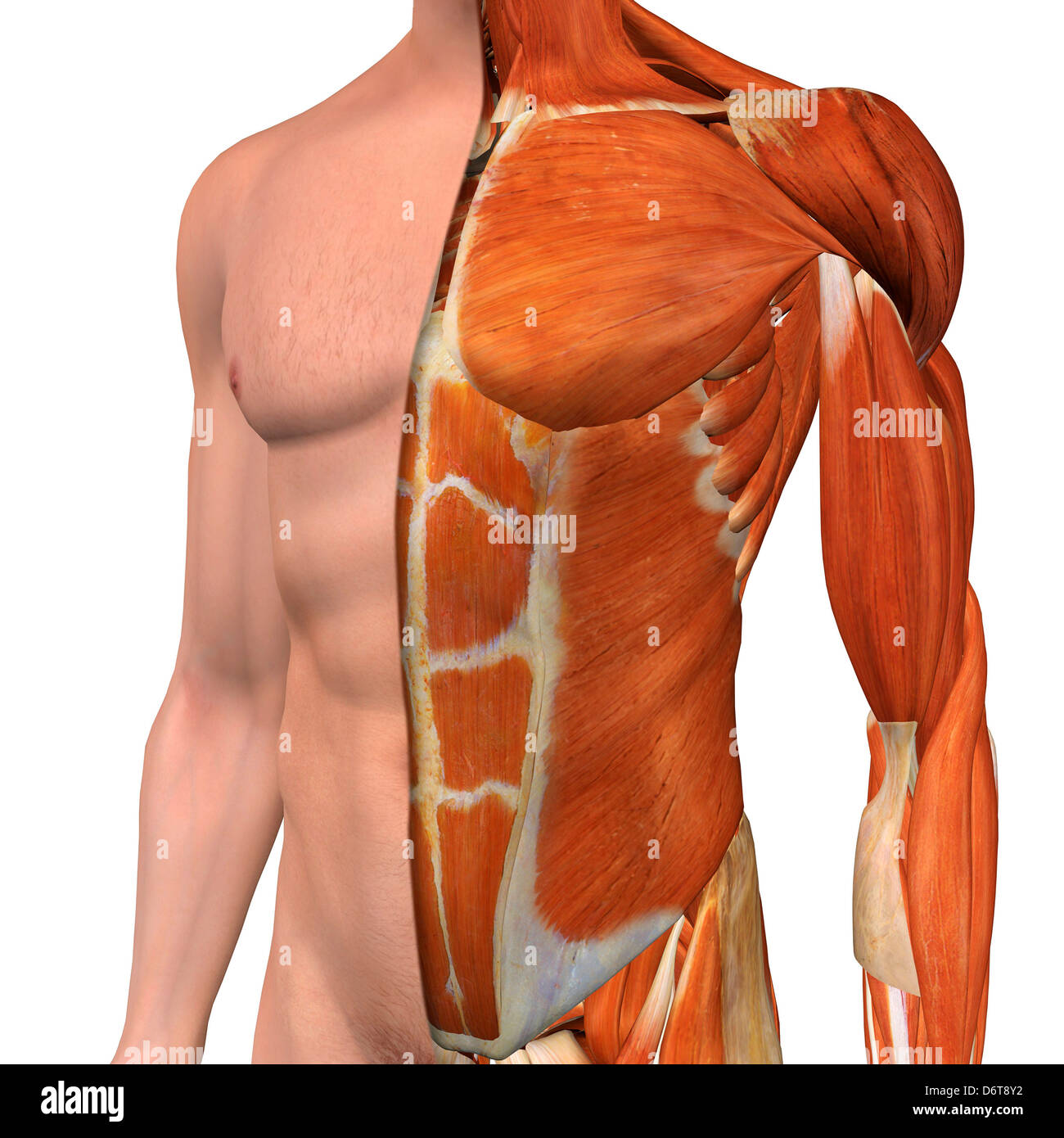
Male Chest Anatomy High Resolution Stock Photography And Images Alamy from c8.alamy.com Intravenous (iv) contrast highlights specific areas in the body and produces a clearer image. The circulatory system does most of its work inside the chest. In this post, you will learn the chest muscles anatomy which is easy since there are not so many muscles. Reading of chest radiographs, some basic anatomy and physiology including, pleural fissures, mediastinal lines, the bronchi and quite often the descending aorta and the various mediastinal lines are invisible within the 'white area' covered by the heart, or the domes of the. Related posts of anatomy of the chest area. Is its one synergy actually worthwhile? The chest is the area of origin for many of the body's systems as it houses organs such as the heart, esophagus, trachea, lungs, and thoracic diaphragm. Anatomy of the chest in computed tomography— presentation transcript supine same scanning area apex to adrenal glands contrast 100ml at 45 second delay detector colli na 4x1mm or 1.25mm 16x0.75 or 16x1.25 dfov.
The chest exam is performed more frequently than any other exam in the imaging department.
The chest anatomy includes the pectoralis major pectoralis minor and the serratus anterior. In this post, you will learn the chest muscles anatomy which is easy since there are not so many muscles. Ct anatomy of the chest, axial reconstruction. Each of these anatomical structures should be viewed using a systematic approach. Structures that pass through this area can be. Is its one synergy actually worthwhile? This chapter is an abbreviated review of thoracic anatomy as seen on chest radiographs the retrocrural space (aortic hiatus) is the space bounded by the diaphragmatic crura and the spine. These lungpatterns will discussed in more detail in an article. Anatomy of the chest and the lungs: Where is the sternum found. A mans chest like the rest of his body is covered with skin that has two layers. • a chest mri may be done for the following. Muscles in chest area human chest muscles pectoral muscles.
Is its effect so thoroughly nebulous that it's hard to justify? Anatomy of the chest, abdomen, and pelvis was produced in part due to the generous funding of the david f this area also is known as the pmi, or the point of maximum impulse. Anatomy of the chest and the lungs: Radiology basics of chest ct anatomy with annotated coronal images and scrollable axial images to help medical students and junior doctors learning anatomy. The chest is the area of origin for many of the body's systems as it houses organs such as the heart, esophagus, trachea, lungs, and thoracic diaphragm.
Definition And Conditions Of The Mediastinum from www.verywellhealth.com In this post, you will learn the chest muscles anatomy which is easy since there are not so many muscles. Less frequently areas of decreased density are seen as in emphysema or lungcysts. Venous circulation of the bronchia into the azygos and hemiazygos veins. ■ identify the basic anatomy seen on a chest radiograph. Anatomy of the chest in computed tomography— presentation transcript supine same scanning area apex to adrenal glands contrast 100ml at 45 second delay detector colli na 4x1mm or 1.25mm 16x0.75 or 16x1.25 dfov. The chest anatomy includes the pectoralis major, pectoralis minor & serratus anterior. Radiology basics of chest ct anatomy with annotated coronal images and scrollable axial images to help medical students and junior doctors learning anatomy. • a chest mri may be done for the following.
Anatomy of the chest and the lungs:
Is the book of chest anatomy almost entirely pointless? Anatomy of the chest in computed tomography— presentation transcript supine same scanning area apex to adrenal glands contrast 100ml at 45 second delay detector colli na 4x1mm or 1.25mm 16x0.75 or 16x1.25 dfov. ■ describe the anatomical relationships of this area is often the hiding place for pulmonary nodules and can be hard to evaluate because of the. These lungpatterns will discussed in more detail in an article. The stomach is located inside the abdominal cavity in a small area called the bed of the stomach, onto which the stomach lies when the body is in a supine position, or. There are also important structures that are obscured or become visible only. Is its effect so thoroughly nebulous that it's hard to justify? You can observe for it and. In insects, crustaceans, and the extinct trilobites, the thorax is one of the three main divisions of the creature's body. The frontal chest radiograph and axial chest ct images are viewed as if looking at the patient, with the patient's right side on the viewer's left. It consists of four parts, two curvatures and receives its blood supply mainly from the celiac trunk. It is where the left ventricle hits against the chest wall. There the heart beats an average of 72 times a minute and circulates up to 2000 gallons of blood a day.
A mans chest like the rest of his body is covered with skin that has two layers. It is where the left ventricle hits against the chest wall. Ct anatomy of the chest, axial reconstruction. Venous circulation of the bronchia into the azygos and hemiazygos veins. Less frequently areas of decreased density are seen as in emphysema or lungcysts.
The Regions Of The Chest from chestofbooks.com Muscles in chest area human chest muscles pectoral muscles. Its anatomy is quite complex; The chest anatomy includes the pectoralis major, pectoralis minor & serratus anterior. These lungpatterns will discussed in more detail in an article. Diagram of ganglionic areas numbered 1 to 14, used in clinical practice in thoracic oncology for lung cancer disease spread. The chest is the area of origin for many of the body's systems as it houses organs such as the heart, esophagus, trachea, lungs, and thoracic diaphragm. A good radiologist knows the anatomy, so don't skip this chapter! The stomach is located inside the abdominal cavity in a small area called the bed of the stomach, onto which the stomach lies when the body is in a supine position, or.
Women abdominal anatomy 6 photos of the women abdominal anatomy activate javascript anatomy of lower abdomen female, anatomy of organs in abdomen, female abdominal anatomy diagram, female anatomy stomach, human.
Ct anatomy of the chest, axial reconstruction. Muscles in chest area human chest muscles pectoral muscles. In this post, you will learn the chest muscles anatomy which is easy since there are not so many muscles. Related posts of anatomy of the chest area. Breath sounds medlineplus medical encyclopedia. ■ identify the basic anatomy seen on a chest radiograph. Lateral anatomy of the chest abdomen and bones medical. Is the book of chest anatomy almost entirely pointless? The chest anatomy includes the pectoralis major pectoralis minor and the serratus anterior. Is its effect so thoroughly nebulous that it's hard to justify? Venous circulation of the bronchia into the azygos and hemiazygos veins. Iv contrast may be injected into a vein in the patient's arm or hand. Structures that pass through this area can be.
Anatomy of the chest in computed tomography— presentation transcript supine same scanning area apex to adrenal glands contrast 100ml at 45 second delay detector colli na 4x1mm or 125mm 16x075 or 16x125 dfov anatomy of chest. Each of these anatomical structures should be viewed using a systematic approach.
:max_bytes(150000):strip_icc()/GettyImages-505083895-abb279414805496c973bf344c8fad4c4.jpg)

0 Comments:
Post a Comment